Stanford Study: Human Brain Grows From Two Separate Origins
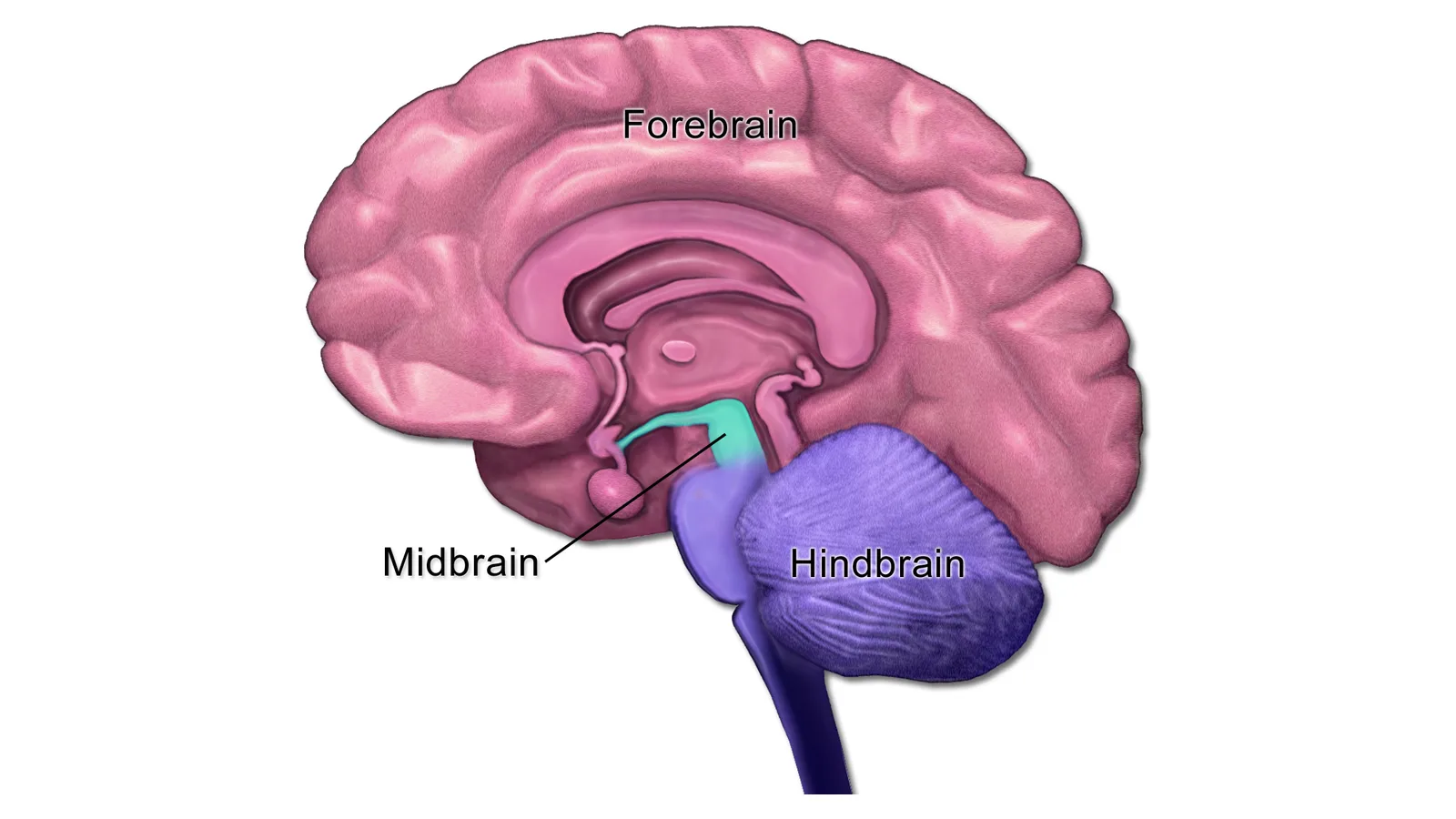
A Stanford Medicine-led team reported on September 18 in the journal Nature Neuroscience that what anatomy textbooks treat as a single organ is built from two independent starting points. Studying mouse embryos during gastrulation — the stage at which the body plan first takes shape — the researchers identified two progenitor cell populations that never mix. One, marked by a gene called Otx2, goes on to form the forebrain and midbrain. The other, marked by Gbx2, is committed to the hindbrain: the brain-stem region that regulates breathing, heartbeat and sleep and controls the face, tongue and throat muscles.
The two populations are mutually exclusive from the earliest stages, and their chromatin — the DNA packaging that decides which genes a cell can reach — is configured so differently that each progenitor is locked into its fate. "We've shown for the first time that the front of the brain arises from a totally different progenitor cell than the back of the brain," said Kyle Loh, PhD, associate professor of developmental biology and the study's senior author. That contradicts the model most of the field has worked from for decades, in which one progenitor cell gives rise to the entire brain. Graduate students Carolyn Dundes and Rayyan Jokhai are co-first authors.
The finding also explains a long-running laboratory failure. Scientists have spent decades unable to generate human hindbrain neurons in a dish. "Previous attempts to make hindbrain neurons likely tried to coax forebrain and midbrain progenitors into hindbrain cells, which our study shows is not possible," Jokhai said. Starting instead from the correct lineage, the researchers turned human pluripotent stem cells into functional hindbrain motor neurons for the first time. According to Stanford Medicine, those cells fired action potentials and made proteins specific to the hindbrain segments that control facial and swallowing muscles.
That is why the result reaches beyond developmental biology. Spinal muscular atrophy is a leading genetic cause of death in children under 1; ALS is usually diagnosed between the ages of 40 and 70. In both, certain hindbrain neurons gradually stop working and patients lose the ability to swallow and eventually to breathe. Because brain-stem tissue cannot be taken from living patients, these diseases have been close to impossible to study directly. What the Stanford team has produced is a model system for that research — not a therapy.
The group also traced the same two-origin pattern back across 550 million years of evolution, finding it in chickens, zebrafish and acorn worms. "Our research suggests that evolution took two existing neural systems and pushed them together spatially," Loh said. Open questions remain: where the spinal cord's own developmental origin lies, and exactly how spinal muscular atrophy and ALS damage hindbrain neurons — the two problems the group says it will take up next.
Sources
- Stanford Medicine NewsPrimary source
- Nature NeurosciencePrimary source
- Earth.comSecondary
Related
First Diplodocus Found Outside North America, in Spain
1,200 Gravity Readings Revive King Tut Hidden-Chamber Debate
Northwestern Study: Disorder Can Make Networks Stabler
Sakurai's Object Is Six Times Hotter Than 30 Years Ago
Leopardus Tilcayo: First New Wild Cat Named in 100 Years
JWST Data Reveals Three Repeating Weather States 20 Light-Years Away
Humpback Filmed Staying With Stillborn Calf, a Species First
Dinosaur Dung Preserves First Known Feathers of a Diving Bird
Trending now
- Universal, Sony Sue Suno Again Over 60,202 Recordings
- China's CXMT Starts Mass Production of 11.95nm G5 DRAM
- Alibaba Open-Sources CT AI That Flags 146 Conditions
- First Diplodocus Found Outside North America, in Spain
- Plugin4Shell: 4 AI Coding Agents Hit, 2 Still Unpatched
- 1,200 Gravity Readings Revive King Tut Hidden-Chamber Debate
- $100M of TikTok's Privacy Deal in Doubt as Judge Keeps 2019 Order
- Gane Named Undisputed UFC Champ, Faces Hokit at MSG Nov. 14
Comments
No comments yet. Be the first.